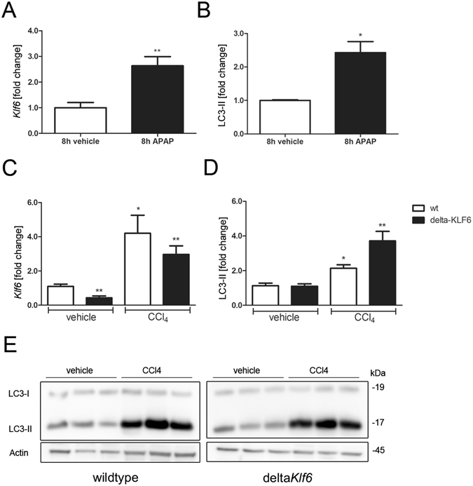
Figure 3.

KLF6 is induced in different experimental models of acute hepatocellular injury. Klf6-expression levels were quantified by qRT-PCR in liver tissue of mice that received vehicle or APAP-injection (A; 500 mg/kg bodyweight after 8 h, n = 6) and in mice that received a single dose of CCl4 (C; 2 µl/g bodyweight, n = 4 for vehicle control and n = 5 for CCl4 treated animals). Autophagy was assessed by LC3 Western blotting and quantified by densitometry of specific protein bands in liver tissue of wildtype mice following APAP-injection (B; Western blot images of APAP-treated mice are show in Supplementary Figure S3) or in wildtype and deltaKlf6 animals after CCl4 injection (D,E). Shown are representative Western blot images of CCl4 treated animals (E; full length Western blot images are given in Supplementary information) and densitometric quantification of LC3-II bands normalized to loading control Actin (E); fold change versus control shown as mean ± SEM of n = 4–6 mice).
