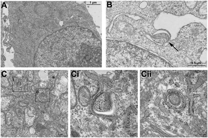
Figure 5.

Autophagosome formation in KLF6 over-expressing HepG2 cells. Under a transmission electron microscope, the autophagosome formation was observed and imaged in HepG2 cells transfected with the empty vector (pcIneo) (A), in HepG2 cells transfected with pcIneo treated with 15 µM rapamycin for 6 h to stimulate autophagosome formation (B) and in KLF6-over-expressing HepG2 cells (pcIneo-KLF6) (C). Representative slides and blow-ups shown are shown of n = 2 independent cell culture experiments.
