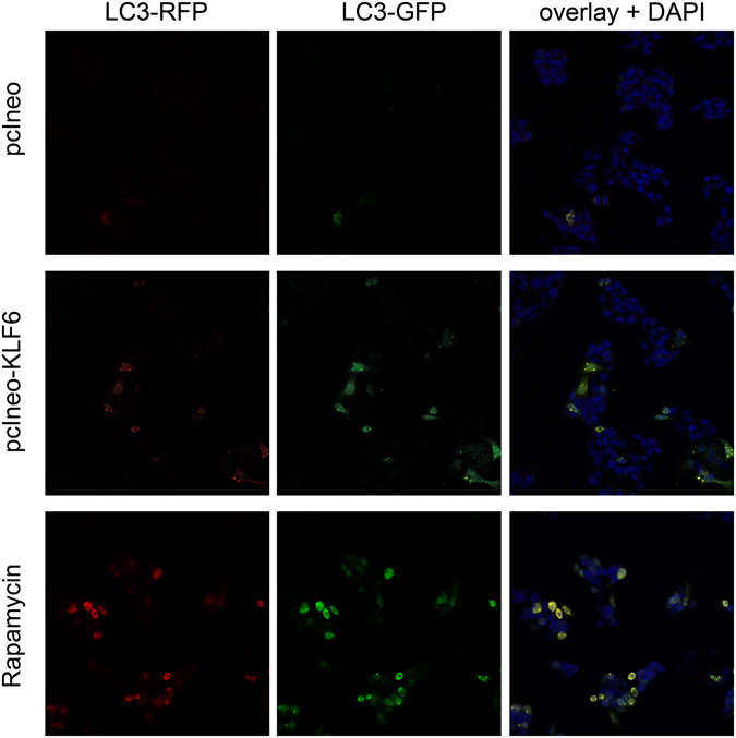
Figure 6.

Autophagosome formation in KLF6 over-expressing HepG2 cells. To visualize formation of autophagosomes in HepG2 cells transfected with empty control vector (pcIneo) or in KLF6 over-expressing HepG2 cells (pcIneoKLF6) we treated the cells with Autophagy Tandem Sensor RFP-GFP-LC3B. As a positive control, we stimulated autophagosome formation via treatment with 15 µM rapamycin for 6 h. Cells were viewed and imaged with a Leica SP8 confocal microscope (20-fold magnification); shown are representative images of n = 3 individual cell culture experiments.
