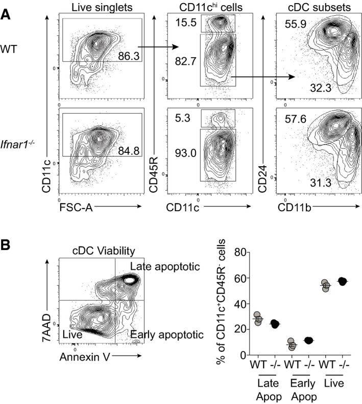
Figure EV2. Ifnar1 −/− FLDCs display normal development and viability.

- FLDCs were cultured for 18 h in medium alone and analyzed for subsets by flow cytometry.
- Following 18‐h culture, FLDCs were stained with 7AAD and Annexin V to assess viability of cDC populations. 7AAD+ Annexin V+ were classed as late apoptotic, 7AAD− Annexin V+ as early apoptotic, and 7AAD− Annexin V− as live cells. Results are mean ± SEM.
