
Figure 2. Ccn1 is highly expressed in stiff regions of orthotopic E0771 tumors.
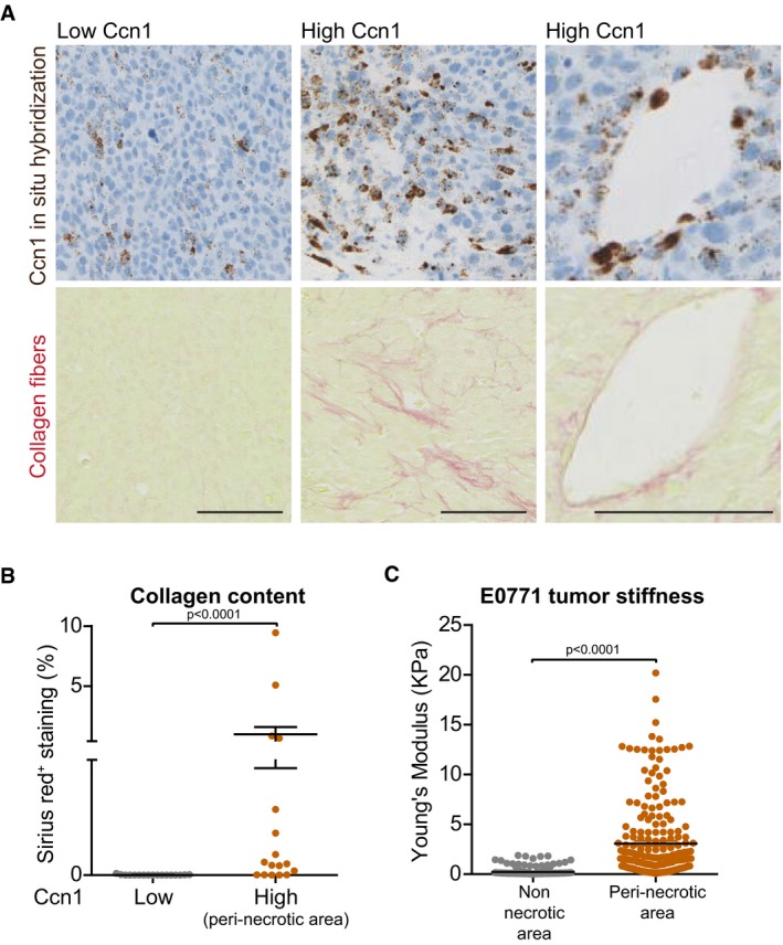
- Representative image of in situ hybridization for Ccn1 mRNA and collagen I and III fibers (Sirius red) of E0771 orthotopic tumors performed on consecutive sections showing that there are tumor regions with high and low expression of Ccn1. The right panels show a blood vessel positive for Ccn1 staining. Scale bar = 100 μm.
- Quantification of collagen I and III fiber content based on Sirius red staining (% of the measured area) showing that higher amounts of collagen fibers are found in regions of the tumor expressing high Ccn1 levels.
- Quantification of tumor stiffness performed by atomic force microscopy showing that higher stiffness is measured in peri‐necrotic regions (bona fide expressing high Ccn1 levels) of the tumor.