
Figure 7. Reduced Ccn1 levels in endothelial cells inhibit cancer cell transendothelial migration.
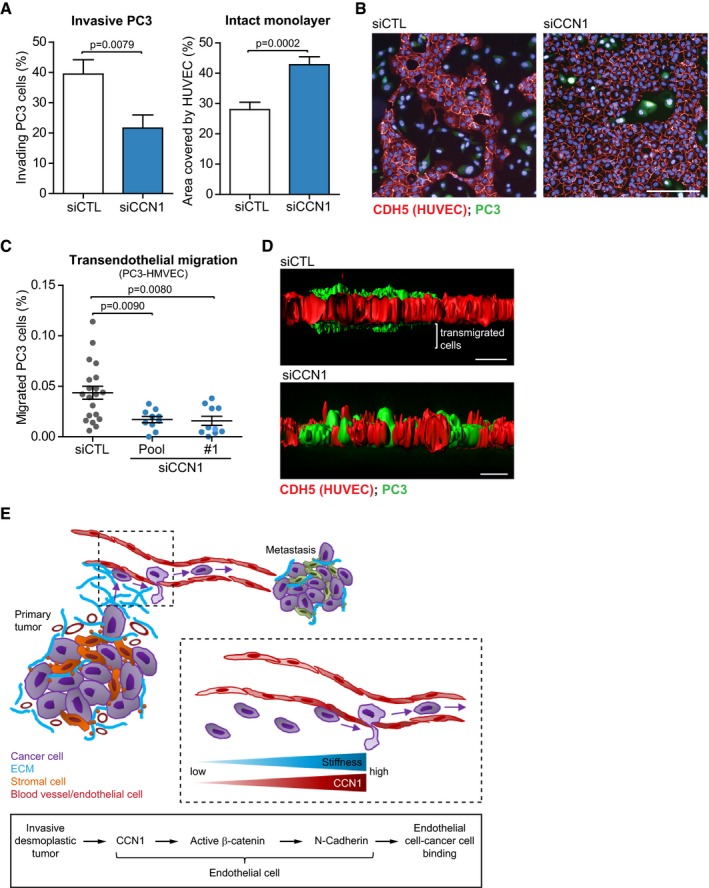
- Silencing CCN1 in a monolayer of HUVECs inhibits the capability of PC3 cells to invade the endothelial monolayer, as measured by reduced number of PC3 cells that invaded the monolayer after 24 h of co‐culture (left), and reduced amount of disrupted regions of the monolayer (right). siCTL n = 21, siCCN1 n = 23 fields assessed from one representative experiment of two.
- Representative immunofluorescence staining of (A). Red = VE‐cadherin (CDH5, HUVECs); green = fluorescently labeled PC3 cells. Scale bar = 100 μm.
- Transendothelial migration assay performed with PC3 cells (clone TEM4‐18) and HMVECs on transwells showing that silencing of CCN1 in HMVECs reduces the passage of cancer cells through the endothelial monolayer. N = 20 (siCTL), 9 (siCCN1 pool) and 10 (siCCN1 #1) fields assessed from two independent experiments.
- Representative immunofluorescence staining of (C). Red = VE‐cadherin (CDH5, HUVECs); green = fluorescently labeled PC3 cells. Scale bar = 20 μm.
- Working model for CCN1 in the tumor vasculature.