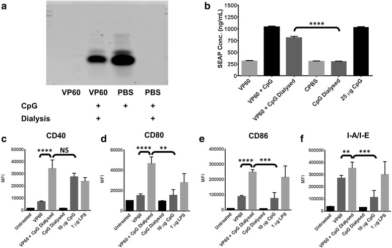
Fig. 3.

Association of CpG with RHDV VLP. The ability of RHDV VLP to naturally associate with CpG was investigated by dialysis with 1 MDa tubing. a CpGs were detected by TBE acrylamide gel electrophoresis stained with Gel Green following dialysis. b Quanti-blue assay on supernatants detecting SEAP secreted from the reporter cell line, RAW-blue, following treatment with CpG associated with VLP. This treatment was repeated with murine BMDCs, detecting the median fluorescence intensity (MFI) of the activation markers c CD40, d CD80, e CD86 and f I-A/I-E by flow cytometry. Results are representative of two independent repeats. L = Hyperladder V, Dial = Dialysed. Statistical analysis performed using unpaired t-tests. NS = Non-significant, ** p < 0.01, *** p < 0.001, **** p < 0.0001
