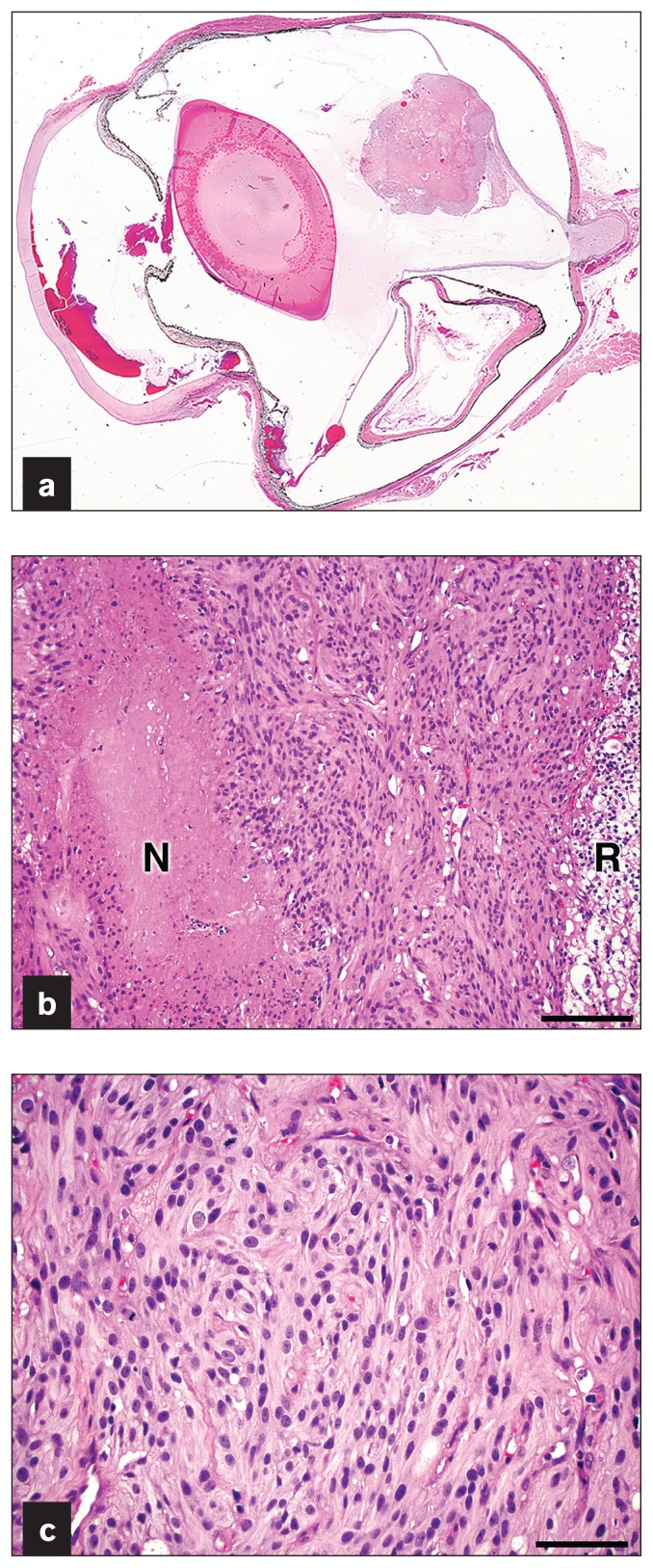
Figure 3.
a — Subgrossly there is a well-circumscribed nodular mass arising from the detached dorsal retina. b — The mass is highly cellular and composed of spindle cells arranged in interlacing fascicles with frequent small caliber vessels and foci of necrosis (“N”) with pseudopalisading of tumor cells. Note that the retina (“R”) contiguous to the neoplasm was degenerate with vacuolar changes. H&E stain. Bar = 100 μm. c — Neoplastic cells have variably distinct cell borders, abundant fibrillar cytoplasm, and oval nuclei with finely stippled chromatin and 1 to 2 variably distinct nucleoli. Mitoses are observed. H&E stain. Bar = 50 μm.

