Fig. 2.

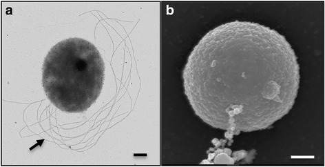
Morphological features. Transmission (a) and scanning (b) electron micrographs of a cell of strain Hulk grown with formate as the electron donor and Fe(III) citrate and sulfate as the electron acceptors, respectively. Arrow in (a) points at the lophotrichous flagella and in (b) at membrane vesicles. Scale bars, 200 nm
