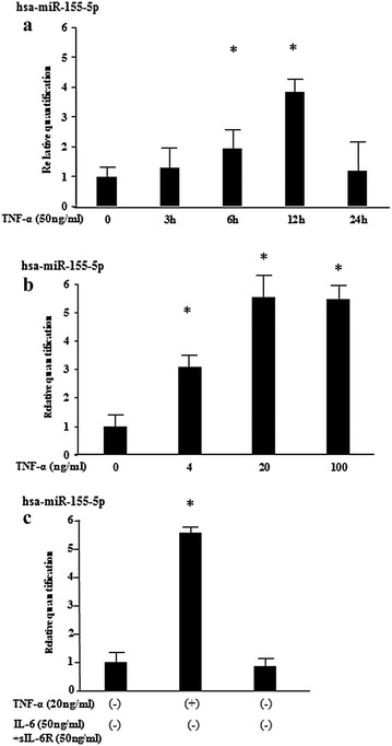
Fig. 2.

TNF-α induces miR-155 expression in RASFs. a RASFs were stimulated with TNF-α (50 ng/ml) for the indicated periods and relative expression of miR-155 were analyzed by qRT-PCR (n = 3). Values represent the mean ± SD of three independent experiments. *p < 0.005 as compared with the value in unstimulated cells. Three experiments were performed using RASFs isolated from three different RA patients and a representative result is shown. b RASFs were stimulated with various concentrations of TNF-α for 12 h and relative expression of miR-155 were analyzed by qRT-PCR (n = 3). Values represent the mean ± SD of three independent experiments. *p < 0.005 as compared with the value in unstimulated cells. Three experiments were performed using RASFs isolated from three different RA patients and a representative result is shown. c RASFs were stimulated with IL-6 (50 ng/ml) plus sIL-6R (50 ng/ml) or TNF-α (50 ng/ml) for 12 h and relative expression of miR-155 were analyzed by qRT-PCR (n = 3). Values represent the mean ± SD of three independent experiments. *p < 0.001 as compared with the value in unstimulated cells. Three experiments were performed using RASFs isolated from three different RA patients and a representative result is shown
