. 2015 Aug 11;16(2):67–72. doi: 10.7181/acfs.2015.16.2.67

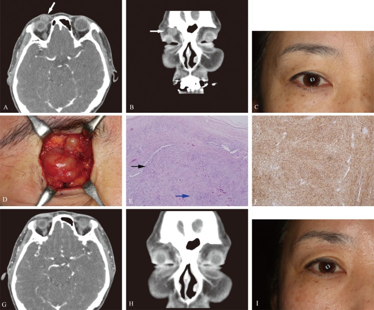
Fig. 1. Case 1. This 51-year-old woman presented with an eight-year history of a progressively enlarging mass on her right upper eyelid (white arrows). (A) Preoperative axial computed tomography (CT) image. (B) Preoperative coronal CT image. (C) Preoperative photograph. (D) Intraoperative photograph showing the mass. (E) Photomicrograph of a tumor specimen showing a biphasic pattern of Antoni A areas (black arrow) with Verocay bodies and Antoni B areas (blue arrow) (H&E, ×40). (F) Immunohistochemical analysis showing positive staining for S-100 protein (S-100, ×100). (G) Postoperative axial CT image. (H) Postoperative coronal CT image. (I) Follow-up photograph at five months.

Fig. 1