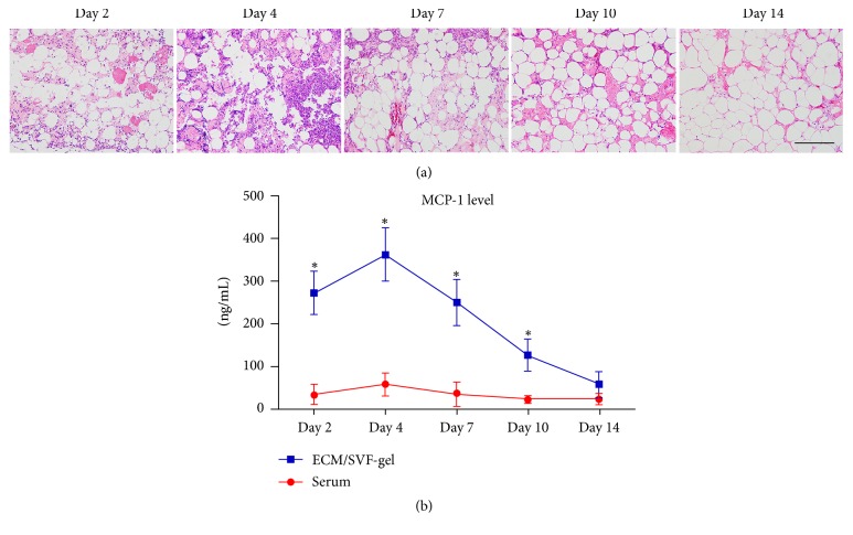
Figure 8.
HE staining of ECM/SVF-gel and MCP-1 expression. (a) Inflammatory cell infiltration was observed obviously in ECM/SVF-gel at an early stage and declined at a late stage of wound healing. Scale bar = 200 μm. (b) MCP-1 expression in ECM/SVF-gel and serum at different time points (∗P < 0.05).

