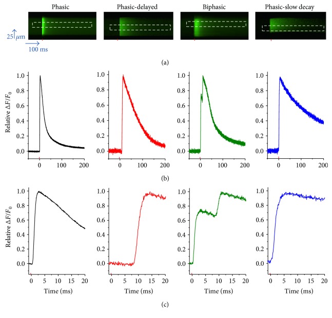
Figure 1.
Muscle fibers cultured for 5 days exhibit multiple patterns of action potential (AP)-induced Ca2+ transients: control conditions. (a) Representative confocal line-scan images of AP-induced Ca2+ transients in 5-day-old cultured muscle fibers maintained in control medium. Note that four different patterns were identified: phasic, phasic-delayed, biphasic, and phasic-slow-decay. The red mark indicates the time when field electrical stimulus was applied, and the dashed rectangle illustrates the region of interest used to measure the time course of the Ca2+ transient. (b) Time course of the AP-induced Ca2+ transients shown in (a). (c) Zoomed-in versions of AP-induced Ca2+ transients shown in (b).

