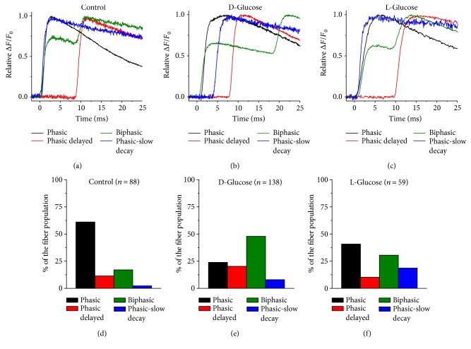
Figure 2.
Sustained elevation of extracellular D-glucose modifies the distribution of AP-induced Ca2+ transients observed in 5-day-old cultured fibers. Zoomed-in and overlapped version of AP-induced Ca2+ transients for control (a), D-glucose (b), and L-glucose (c) challenged fibers. (d–f) Summary of distribution of AP-induced Ca2+ transients for fibers exposed to control isotonic medium (d), D-glucose (e), and L-glucose (f). Fibers exposed to D-glucose displayed a significantly larger proportion of biphasic action potential-induced Ca2+ transients when compared to control counterparts (X2, n = 286, p value < 0.05).

