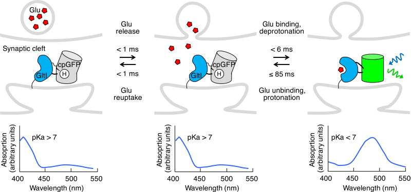
Figure 2.
Genetically encoded transmitter indicators (GETIs). iGluSnFR reports glutamate with increased fluorescence (above). A glutamate-induced conformational change in the glutamate-binding domain from a bacterial glutamate transporter (Glt1) induces loss of the proton from the chromophore, shifting its absorbance peak to ~490 nm (below) and allowing excitation by 488-nm light (blue sinusoidal arrow), with resulting green emission (green sinusoidal arrow). Binding time was measured in vitro for an increase in glutamate concentration from 0 to 4.6 µM (ref. 46). The indicated unbinding time is an upper limit deduced from live cell experiments.

