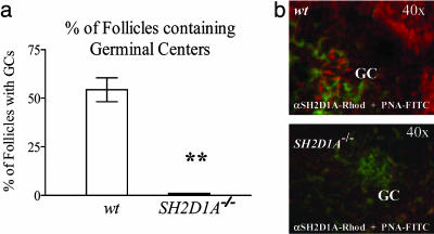
Fig. 2.
Follicular GC formation. (a) Reduced number of GC-containing follicles in the spleens of SH2D1A-/- mice. The number of GC-containing follicles was determined from at least three different consecutive stained cryosections taken from the spleens of the TNP-KLH mice, described in Fig. 1c. B cell follicles were identified by anti-CD45R/B220-phycoerythrin, and GCs were stained with PNA-FITC. y axis, percentage of follicles containing one or more GCs. Filled bar, SH2D1A-/- BALB/c; open bar, wt BALB/c(**, P < 0.001; n = 4; n = number of mice tested per experiment). Results are representative of three independent experiments. (b) Expression of the SH2D1A protein in follicular GCs. Cryosections of the spleen of wt BALB/c mice 12 days after immunization with NP-KLH were stained with immunofluorescent antibodies, as indicated. Fluorescence was recorded in a Nikon fluorescent microscope. To detect SH2D1A, acetone-fixed and permeabilized spleen sections were first stained with a rabbit anti-mouse anti-SH2D1A antibody (SH2D1A). In a second step, the sections were stained with Rhodamine-labeled donkey anti-rabbit IgG (red). Shown are FITC-labeled-PNA- (green) identified GCs. (Lower) One of the few GCs identified on spleens of SH2D1A-/- injected mice. Antibodies used for staining are indicated at the bottom; magnification is indicated at top right.

