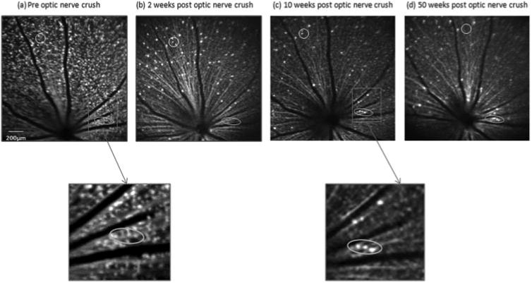
Figure 2.

In vivo bCSLO images in the same retinal area of a transgenic Thy-1 CFP mouse before (a) and 2 (b), 10 (c), and 50 (d) weeks after optic nerve crush. Progressive loss of fluorescent spots is illustrated. A cluster of Thy-1 CFP–expressing RGCs (circled) demonstrated progressive loss of Thy-1 CFP after optic nerve crush. A few cells were diminished in fluorescence at week 2 but returned their signals at 10 and 50 weeks after crush (ovals). The reappearance of fluorescence likely signified the return of normal expression of Thy-1 after the stress response.
