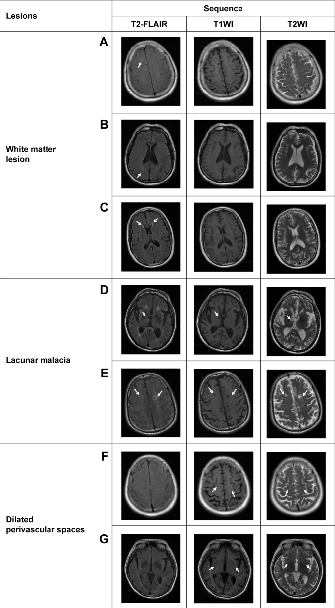
Figure 1.
Examples of the characteristic changes shown on T2-FLAIR in comparison with those shown on T1WI and T2WI.
Notes: T2-FLAIR is comparatively more sensitive in detecting white matter lesions as shown in subcortical white matter (A), deep white matter (B), and periventricular white matter (C). Lacunar malacia in multiple regions are also seen more clearly on T2-FLAIR (D, E), shown as lower signal intensity in the central part surrounded by high signal intensity. In contrast, compared to T1WI and T2WI, the dilated perivascular spaces located in multiple sites are not seen on T2FLAIR, for example, in the subcortical region (F), and basal ganglia and surrounding region (G).
Abbreviations: T1WI, T1-weighted image; T2WI, T2-weighted image; T2-FLAIR, T2-weighted fluid-attenuated inversion recovery.

