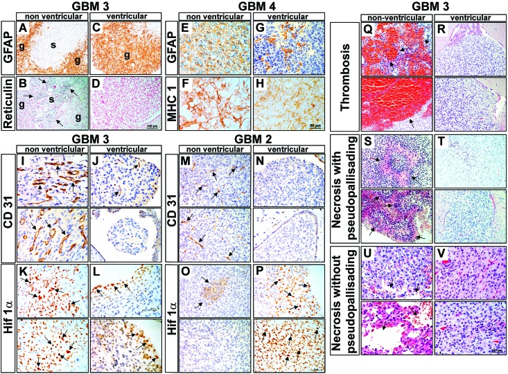
Figure 4.
Morphologic features and immunohistologic markers are distinct between ventricular and nonventricular xenograft tumors. (A) GFAP expression and (B) reticulin staining (arrows; adjacent section) in GBM3 PDOX show the biphasic glial (g) and sarcomatous (s) components that characterize the glioblastoma-variant gliosarcoma. The gliosarcoma biphasic staining pattern is suppressed in ventricular tumors, as demonstrated by (C) the loss of GFAP-negative regions and an absence of reticulin staining, thus indicating that (D) only the glial (g) component is present. (E) GFAP is highly expressed in GBM4 nonventricular tumors. (F) An adjacent tissue section is stained with the human marker MHCI. (G) GFAP expression is restricted to cell clusters in the ventricular tumor. MHCI staining reveals human tumor cells throughout the GFAP-negative regions in the adjacent section, indicating that the absence of GFAP staining is not due to a lack of human tumor cells (H). (I) CD31 staining of GBM3 tumors reveals numerous hyperplastic blood vessels (arrows) in nonventricular tumors, typical of human glioblastoma. In comparison, (J) ventricular tumors display much less vascular hyperplasia, particularly in the small spheroid-like tumor masses floating freely in the ventricle. (K) The Hif1α−positive cells in GBM3 indicate greater hypoxia in nonventricular tumors; (L) ventricular tumors were less hypoxic. As for GBM3 tumors, CD31 staining of GBM2 revealed more vascular hyperplasia in (M) nonventricular tumors relative to (N) ventricular tumors. (O) Focal Hif1α-positive cells were observed in GBM2 tumors (arrows) and (P) a larger area of Hif1α-positive cells (arrows) in ventricular tumors. (Q–V) GBM3. Thrombosis was (Q) frequent (arrows) in nonventricular tumors but was (R) absent from ventricular tumors. PP necrosis was (S) prominent (arrows) in nonventricular tumors but (T) absent in ventricular tumors. Similarly, necrosis without pseudopalisades was (U) frequent (arrows) in nonventricular tumors but (V) absent in nonventricular tumor. Scale bars: D, 100 μm (applies to A–D); H, 50 μm (applies to E–P); and V, 100 μm (applies to Q–V).

