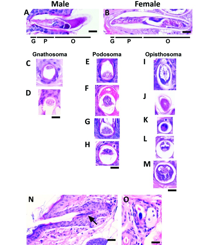
Figure 4.
Longitudinal and transverse sections of mites observed during topographic analysis of TRP1/TCR mouse skin sections. The diversity in appearance of mites is shown with representative images of Demodex mites viewed under high magnification (600×). The body segments in (A) male and (B) female mites include the gnathosoma (mouth parts [G]), podosoma (cranial region with legs [P]), and opisthosoma (caudal region [O]). Male and female mites differ in length and reproductive structures. Transverse sections of the (C and D) gnathosoma, (E–H) podosoma, and (I–M) opisthosoma are shown. Mite eggs (N, longitudinal; O, transverse) appear as dark basophilic structures. Scale bar, 20 μm (A-M, O); 30 μm (N).

