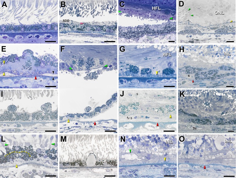
Figure 1.
Fifteen phenotypes of retinal pigment epithelial cell morphology in advanced age-related macular degeneration. Assembled from previous descriptions.6,8,13,15,18 Submicrometer epoxy resin sections of OTAP-postfixed specimens were stained with toluidine blue. All scale bars are 20 μm. Abbreviations: BLamD, basal laminar deposits; BLinD, basal linear deposits; ELM, external limiting membrane; HFL, Henle fiber layer; INL, inner nuclear layer; RPE, retinal pigment epithelium. Yellow arrowheads: BLamD; red arrowheads: calcification in Bruch's membrane; green arrowheads: ELM. (A) “Nonuniform” RPE: slightly nonuniform morphology and pigmentation with small patches of early BLamD. (B) “Very Nonuniform” RPE: highly nonuniform in shape and pigmentation; pink arrowhead: melanosomes within apical processes; SDD, subretinal drusenoid deposits. (C) “Vitelliform”: exploded RPE lipofuscin/melanolipofuscin granules are mixed with outer segment debris. Intact RPE is also present in the HFL. (D) “Subducted”: flattened cells with spindle-shaped melanosomes and lipofuscin/melanolipofuscin granules are located between persistent BLamD and Bruch's membrane. Border of atrophy is defined by a curved ELM. (E) “Shedding” RPE: basal translocation of granule aggregates into a thick continuous layer of BLamD; black arrowheads: BLinD. (F) “Intraretinal” RPE: anterior migration through ELM into the retina. Epithelial component remains atop BLamD (bottom). Photoreceptors have degenerated. Loss of soft druse contents and detachment of retina are artifacts. (G) “Dissociated” RPE: fully granulated and nucleated cells in the atrophic area, adherent to BLamD. Some RPE granules are translocated among HFL fibers. (H) “Melanotic”: contain spherical polydisperse melanosomes and are located inside and internal to a sub-RPE fibrous scar(s) without BLamD, encapsulated by a light blue–staining collagenous material. (I) “Sloughed” RPE: spherical, fully granulated, nucleated cells released into the subretinal space. (J) “Entombed” by a subretinal scar (s) and a sub-RPE scar (fv.s). Persistent BLamD divides these compartments. (K) “Entubulated”: nucleated RPE cell within the lumen of outer retinal tubulation, that is, cone photoreceptors surviving RPE degeneration, scrolled up by Müller cells. (L) “Bilaminar”: two layers of fully pigmented cells delimited by dotted line, adherent to BLamD. (M) “Vacuolated” RPE: cells with a single large vacuole delimited by effaced cytoplasm. (N) “Atrophy with BLamD”: absent RPE and persistent BLamD. Photoreceptors have atrophied. Teal arrowhead: ELM delimits end-stage outer retinal tubulation. (O) “Atrophy without BLamD”: absent RPE, absent BLamD, absent photoreceptors. Glial scar and INL contact Bruch's membrane. (A, B, E–G, I, J, L–O) reprinted from Zanzottera EC, Messinger JD, Ach T, Smith RT, Freund KB, Curcio CA. The Project MACULA retinal pigment epithelium grading system for histology and optical coherence tomography in age-related macular degeneration. Invest Ophthalmol Vis Sci. 2015;56:3253–3268. © 2015 ARVO. (C) reprinted from Balaratnasingam C, Messinger JD, Sloan KR, Yannuzzi LA, Freund KB, Curcio CA. Histologic and optical coherence tomographic correlates in drusenoid pigment epithelium detachment in age-related macular degeneration. Ophthalmology. 2017;124:644–656. © 2017 by the American Academy of Ophthalmology. (D, H) reprinted from Zanzottera EC, Messinger JD, Ach T, Smith RT, Curcio CA. Subducted and melanotic cells in advanced age-related macular degeneration are derived from retinal pigment epithelium. Invest Ophthalmol Vis Sci. 2015;56:3269–3278. © 2015 ARVO. (K) reprinted from Schaal KB, Freund KB, Litts KM, Zhang Y, Messinger JD, Curcio CA. Outer retinal tubulation in advanced age-related macular degeneration: optical coherence tomographic findings correspond to histology. Retina. 2015;35:1339–1350. © 2015 by Ophthalmic Communications Society, Inc.

