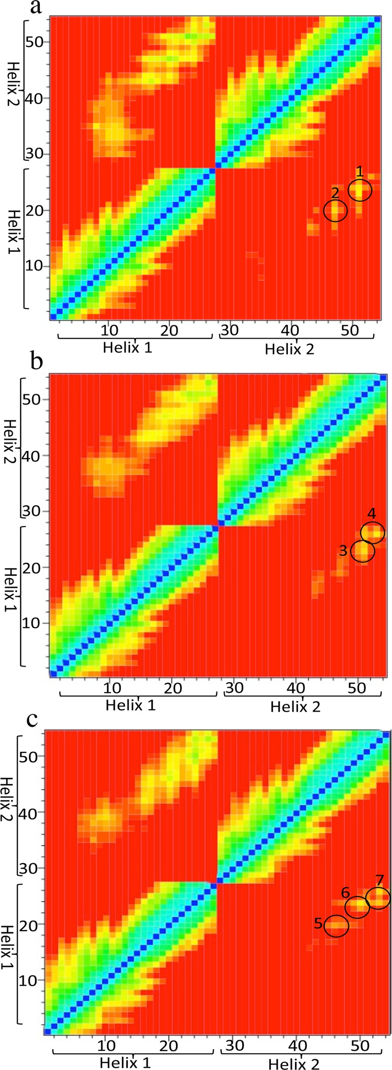
Figure 7.

Contact matrices (heat maps) showing specific interactions between two mutated A2A TM5 helices (“helix 1” and “helix 2”) with the following residues mutated: M177A (a), M1935.54A (b), and M1935.54I (c). Results shown are the average for each ensemble. Interhelical distances at the 15 and 12 Å cutoff distances are shown in the top left and lower right quarter of panels (a–c), respectively. The color scale is as indicated in Figure 6. Circles indicate areas with key interhelical contacts. The identified amino acid interactions are numbered as follows: (1) M1935.54 with M1935.54; (2, 3) V1965.57 with Y1975.58 and Y1975.58 with Y1975.58; (4) Y1975.58 with I2005.61 and R1995.60 with R1995.60; (5) L1925.53 with I1935.54, V1965.57 with Y1975.58, and Y1975.58 with R1995.60, (6) Y1975.58 with I2005.61 and Y1975.58 with R1995.60; and (7) R1995.60 with R1995.60.
