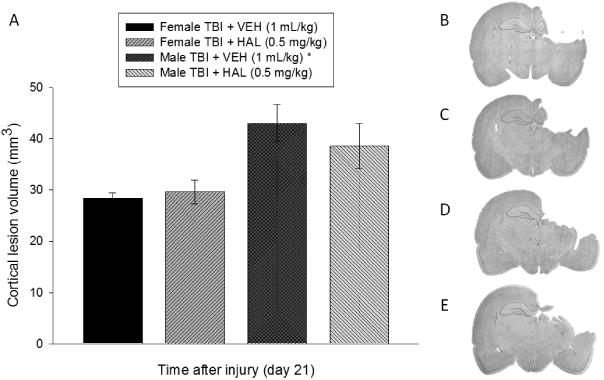
Fig. 5.

A. Mean (± S.E.M.) cortical lesion volume (mm3) 3 weeks after controlled cortical impact injury. B, C, D, and E depict average size lesions, stained with Cresyl violet, at the level of the dorsal hippocampus for Female TBI + VEH, Female TBI + HAL, Male TBI + VEH, and Male TBI + HAL, respectively. * p < 0.05 vs. Female TBI + VEH and Female TBI + HAL. No other comparisons were significant [p > 0.05].
