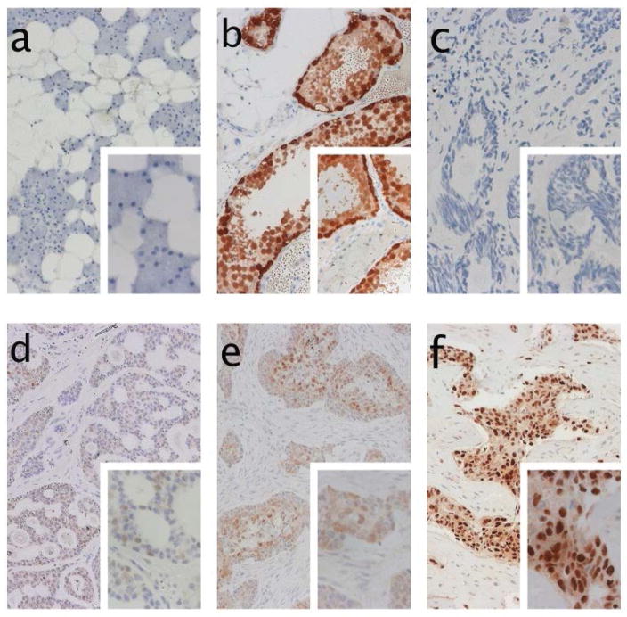
Figure 3.
Immunohistochemical staining of pan-MAGE (M3H67 antibody): a) Negative control of parotid gland tissue. b) positive control staining of testicular tissue. c) negative staining of tumor tissue. d) weakly positive staining of pan-MAGE. e) moderately strong expression of pan-MAGE in tumor tissue with solid growth pattern, stroma tissue is negative. f) example of strong expression of pan-MAGE in >70% of ACC tumor cells.

