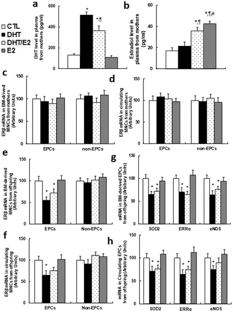
Fig 1. Perinatal testosterone exposure suppresses ERβ expression and its target genes in EPCs in young offspring (2 months old).
2-month old female mice were exposed to 5mg of 60-day release hormone pellets that contained either dihydrotestosterone (DHT) alone, estradiol (E2) alone, combined DHT and E2 (DHT/E2), or controlled vehicle (CTL) during a 7-week perinatal period. The mothers were sacrificed to measure the plasma hormone levels, and the MNCs (including EPCs and non-EPCs) were isolated from either the bone marrow or peripheral blood for analysis of gene expression. The male offspring was also sacrificed at 2 months old for isolation of MNCs (including EPCs and non-EPCs) for further analysis. (a) Dihydrotestosterone (DHT) level in plasma from mothers, n = 8. (b) The estradiol (E2) level in plasma from mothers, n = 8. (c) The ERβ mRNA in BM-derived MNCs from mothers, n = 7. (d) The ERβ mRNA in Circulating MNCs from mothers, n = 7. (e)The ERβ mRNA in BM-derived MNCs from male offspring, n = 6. (f) The ERβ mRNA in Circulating MNCs from male offspring, n = 6. (g) The mRNA levels in BM-derived EPCs from male offspring, n = 7. (h) The mRNA levels in Circulating EPCs from male offspring, n = 7. *, P<0.05, vs CTL group; ¶, P<0.05, vs DHT group; #, P<0.05, vs DHT/E2 group. Results are expressed as mean ± SEM.

