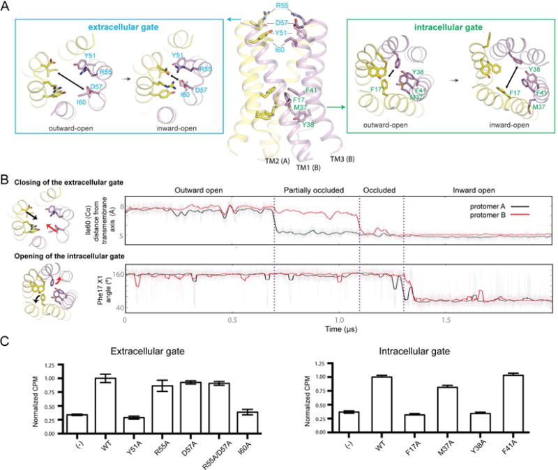
Figure 4. Key molecular events during the outward-open to inward-open transition.

(A) During state transitions, changes to the extracellular gate (top-down view) and the intracellular gate (bottom-up view) are shown in the boxed areas. Two-headed arrows indicate direction of helical movement observed during simulation. (B) Sequential order of events during the outward-open to inward-open transition (Simulation 4). The conformational transition begins when TM3 from each protomer displaces inward, as measured by the distance of the I60 Cα atom to the central axis of the helical bundle (top panel). Inward displacement of TM3 destabilizes packing between TM1 and TM3 in the region around Pro19 (Figure S5). Inward displacement of TM3 on the second protomer occurs within ∼0.4 μs of the first protomer displacing inward, forming a fully occluded state. Transition to the inward-open state is favored by the formation of backbone hydrogen bonds along TM1, which correlates with formation of the kinked conformation of TM1 (Figure S5). These collective motions favor the rotation of Phe17 to move away from the central pore and pack against TM3 (bottom panel). (C) Glucose (14C) uptake activities in E. coli are shown for LbSemiSWEET with alanine point mutations in extracellular (left) and intracellular (right) gates compared to wild type and control (empty vector) (±s.d., n=3). See also Figure S4, S5, S7 and Table S2.
