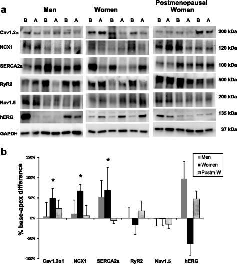
Fig. 1.

Comparison of protein expression between the base (B) and apex (A) of the left ventricular epicardium in humans. Protein samples from the base (B) and the apex (A) of the LV epicardium were obtained from 3 men, 3 women, and 3 postmenopausal women and were probed with antibodies to compare the relative expression of Cav1.2α, NCX1, SERCA2a, RyR2, Nav1.5, and hERG. Panel a shows the protein densities which were normalized with respect to GAPDH. Panel b is a histogram of normalized band intensities expressed as base-apex percent differences derived from an analysis of myocardium from 6 men, 11 women, and 6 postmenopausal women
