. 2017 Jul-Aug;43(4):753–761. doi: 10.1590/S1677-5538.IBJU.2016.0454

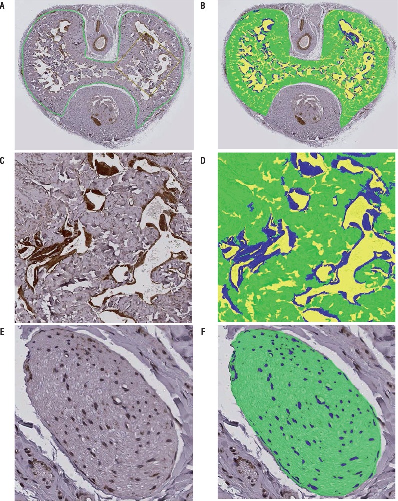
Figure 1. Image analysis method. (A, C) positive immunohistochemical staining of α-smooth muscle actin (dark brown) in a penile midshaft specimen from a control rat. The area of the corpora cavernosa was circled manually in green color in (A). (B, D) software color segmentation performed on the images in (A) and (C) shows the blue-colored smooth muscle and green-colored non-immunoreactive tissue areas that were recognized and captured by the automated digital image analyzer for area measurements, as well as the yellow-colored blank spaces. (E) Positive immunohistochemical staining of nNOS (dark brown) in the dorsal nerve in a penile midshaft specimen from a control rat. (F) software color segmentation performed on the image in (E) shows the blue-colored nNOS and green-colored non-immunoreactive tissue areas that were recognized and captured by the automated digital image analyzer for area measurements.

Figure 1