
Fig. 3.
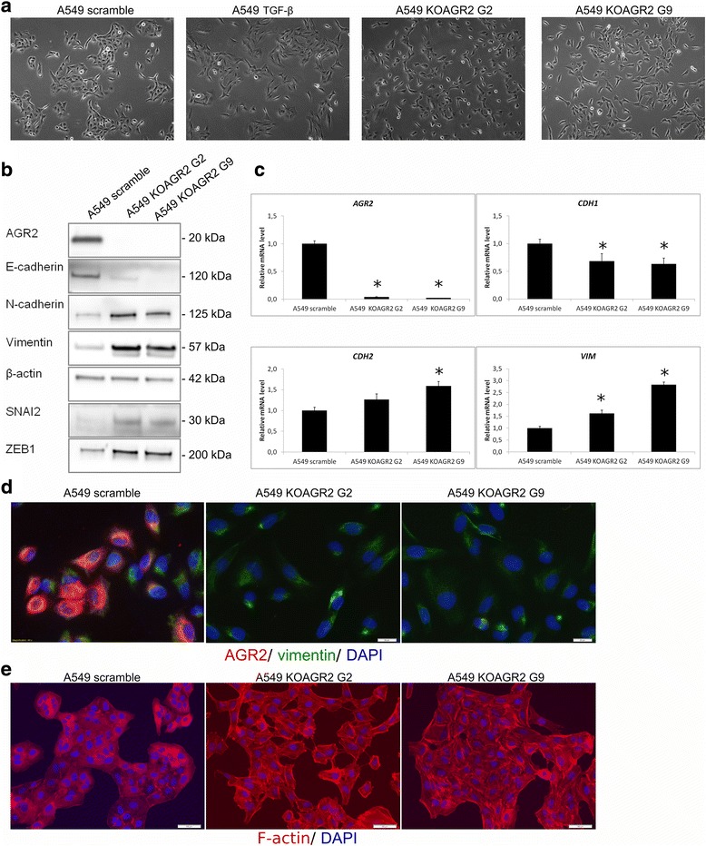
AGR2 loss mimics the changes associated with TGF-β-induced EMT. a A shift in morphology from rounded to spindle-like phenotype of cells was observed for both A549 KOAGR2 cells and A549 cells exposed to TGF-β in comparison with untreated A549 cells. b Protein levels of AGR2 and EMT markers were analyzed in protein lysates from A549 and two clones of A549 KOAGR2 cells named clone G2 and G9. c mRNA levels of AGR2, CDH1, CDH2, VIM were analyzed by RT-qPCR, 18S rRNA served as an endogenous control for data normalization. Data for qPCR are the mean +/− standard deviation obtained from three independent experiments. Statistical significance (P < 0.05) is indicated by asterisks. d Changes in subcellular localization of vimentin in A549 and A549 KOAGR2 were analyzed using immunofluorescence. Staining for AGR2 (red), vimentin (green) and nucleic staining using DAPI was examined. e The A549 and A549 KOAGR2 were also stained with CytoPainter phalloidin-iFluor 594 reagent to detect morphological changes associated with F-actin reorganization. Nuclei were visualised with DAPI