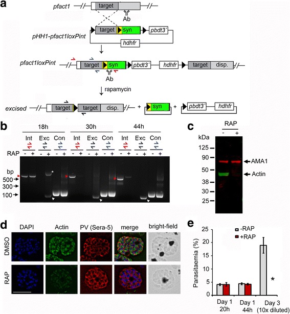
Fig. 1.

Strategy for modification and conditional disruption of pfact1. a Schematic showing single crossover allelic replacement of the endogenous pfact1 gene in the P. falciparum 1G5DiCre line with the construct pHH1-pfact1loxPint, which includes a 493-bp homologous targeting region (target) followed by the loxPint module (yellow) followed by a recodonised pfact1 sequence (syn), coding for the rest of the pfact1 open reading frame. LoxP sites are depicted as black triangles. Integration replaced the pfact1 3´ UTR with that of P. berghei dihydrofolate reductase gene (pbdt). The hdhfr cassette confers resistance to the drug WR99210. Following RAP-induced recombination between loxP sites, the 3´ half of the pfact1 gene, coding for the C-terminal 192 aa residues, was removed along with the introduction of STOP codons. Positions of hybridisation of primers for diagnostic polymerase chain reaction (PCR) have been depicted as red, blue and black half arrows. b Time course of diagnostic PCR with primers showing loss of the integrant pfact1 locus 18, 34 and 44 h post-RAP treatment (Int, red half arrows and red arrow heads), with a concomitant appearance of the excised locus (Exc, black half arrows and white arrow heads) and no reduction in product of the control PCR (Con, blue half arrows). The asterisk (*) marks an intermediate product of excision which is rapidly lost by 34 h. c Western blot showing reactivity to a PfACT1 antibody raised against residues 239 to 253 (Actin) is lost within 44 h post-RAP-induced excision; anti-AMA1 staining is used as control. See also Additional file 1: Figure S1b d IFA shows a loss of reactivity of PfACT1 KOs to the anti-PfACT1 antibody. The PV is depicted in red (Sera-5). Scale bar 5 μm. See also Additional file 2: Figure S2. e Parasitaemia counts indicate an abrupt death of PfACT1 KO parasites (RAP) in the following asexual cycle (day 3). N > 1000. Error bars represent standard deviation (SD)
