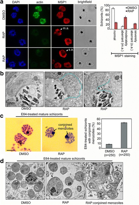
Fig. 3.

PfACT1 is required for normal cytokinesis. a IFA of PfACT1 KO and control schizonts. Aberrant staining of the plasma membrane marker MSP1 (red) depicts dysmorphic merozoites in the schizonts (white arrow) in the absence of PfACT1 (actin). Scale bar 5 μm. Normal, moderately aberrant (m.a.), and severely aberrant (s.a.) MSP1 staining has been exemplified (white arrows) and quantified (right). N > 300, error bars represent SD. b TEM on C1-treated mature schizonts stalled just prior to egress. Whilst medially resident daughter merozoites have distinct, separated membranes apposed to the FV membrane in DMSO controls (black arrows), aberrant membranous pockets including merozoite material are observed in RAP-treated parasites (double black arrows and outlined by blue dotted line). c Giemsa-stained, E64-treated mature schizonts show conjoined merozoites (black arrows) with ~50% frequency in the RAP-treated sample as compared to <10% in DMSO controls. N > 300, scale bar 5 μm. Error bars represent SD. d TEM of E64-trapped merozoites. Merozoites in DMSO controls are distinct and well formed, with organelles not included within the FV membrane (left panel). The RAP-treated population has FVs (black arrow) which include organelles normally resident in daughter merozoites. Some merozoites show aberrant surface architecture (asterisked). Merozoites conjoined to each other are frequently seen in the PfACT1 KO population (right panel). TEM scale bar 500 nm. Other scale bars 5 μm
