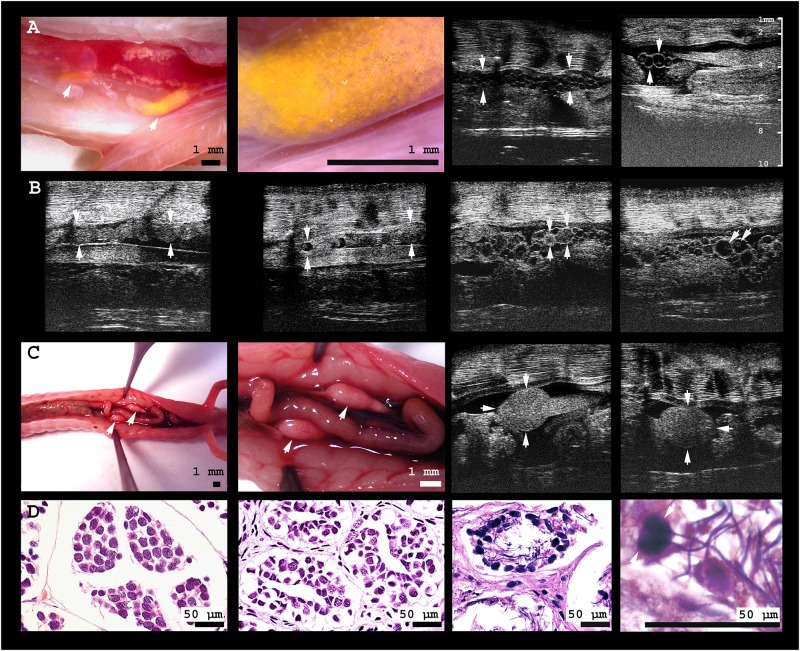
Fig 3. Gonads of male and female olms.
A From left to right: (i) digital microscopic image of the caudal third of the body cavity of a female olm. The yellow pigmented ovary is located adjacent to the kidneys; the intestine is deviated to allow for free vision on the gonads. (ii) In close-up, small spherical follicles are visible within the translucent, comparably inactive ovary. (iii) Ultrasonographic image of an animal with follicles of small to medium size (≤ 0.5 mm), and of (iv) medium size (≤ 0.8 mm), including several homogenously echoic follicles; ovary margins are indicated by arrowheads. B From left to right: (i) The oviduct of the same individual without, and (ii) with oocytes inside; oviduct margins are indicated by arrowheads. (iii) Large oocytes of homogenous intermediate echogenicity, presumably representing sites of recently ovulated follicles (CLs) are indicated by the arrows, adjacent to (iv) comparably large (1.1 mm) vitellogenic follicles inside the ovary of the same individual; the yolk is visible as a slightly echogenic sphere within the anechogenic follicles. Note the large size differences of follicles within the ovary. C From left to right: (i) digital microscopic image of the caudal body cavity of a male olm. Testes are connected to and located cranially of the kidneys, adjacent to the intestine; (ii) close-up of the testes. (iii) Ultrasonographic image of left and (iv) right testis. D From left to right: (i) histological image of an individual with inactive testes, (ii) empty seminiferous tubules, and (iii) active spermatogenesis in the testes of another male olm; (iv) close-up of the testicular spermatozoa.

