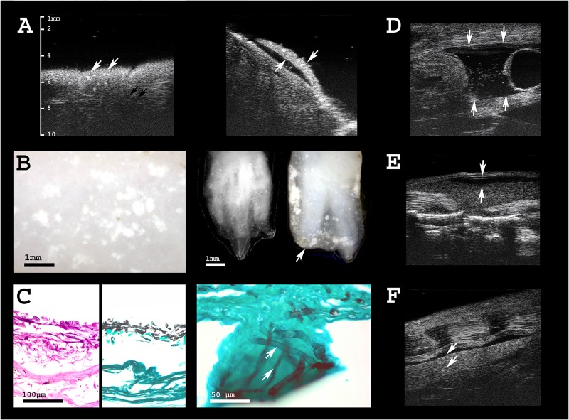
Fig 4. Health assessment of olms.
A Ultrasonographic image of the skin of an olm that died due to Saprolegnia infection. White arrows indicate hyperechogenic foci in the skin that are not visible by adspection of the animal. Black arrows point to circular, hypoechogenic skin mucus glands (left), variably distributed across different body regions. Gills are severely damaged (right). B Digital microscopic images of the same individual; white spots indicate skin lesions caused by the fungal infection (left). Extremities are also severely affected; digits of both, three-toed hand and two-toed foot are partly destroyed by the infection, indicated by the arrow (right). C From left to right: histological sections of the infected skin, whose outer layers are detached and lost their integrity. Grocott stain of infected skin, and close-up to visualize fungal hyphae (stained in black) invading the epidermis. D Free coelomic fluid (physiological in amphibians; arrows) is augmented (ascites) and contains hyperechogenic particles in Saprolegniasis, indicating deteriorated health status. The bladder is seen on the right side of the image as echogenic membrane of 0.1–0.2 mm thickness, surrounding a circular space of anechogenic fluid at the caudal end of the body cavity. E One individual presented with subcutaneous edema, i.e. lymph accumulation within the dorsal lymph sac, with a wide range of possible underlying causes. F Two individuals showed sporadic hyperechogenic spots in the kidney that may indicate parasitic infection or renal deposits.

