Figure 4.

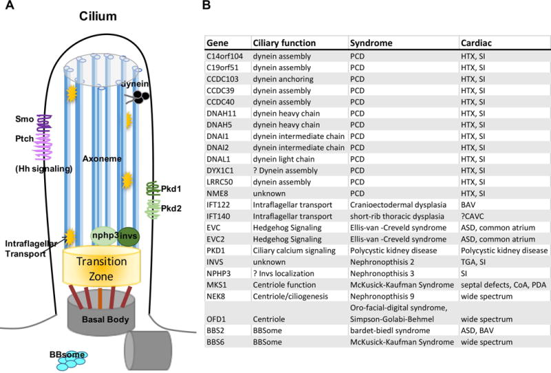
Cilia in CHD A Diagram of a cilium, showing the ciliary axoneme (blue) based on the mother centriole (gray) and linked via the transition zone (orange). B Syndromes and CHD linked to human cilia mutations.

Cilia in CHD A Diagram of a cilium, showing the ciliary axoneme (blue) based on the mother centriole (gray) and linked via the transition zone (orange). B Syndromes and CHD linked to human cilia mutations.