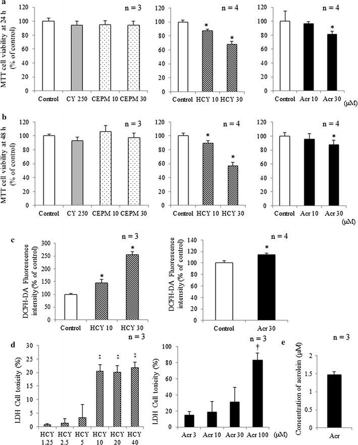
Fig. 2.

Myocardial cytotoxicity induced by HCY or acrolein. H9c2 cell viability after a 24-h and b 48-h exposure to CY alone and CY metabolites was assessed by MTT assay (mean + SD from 3 to 4 independent experiments). c Fluorescence intensities, corresponding to levels of H2O2, in control samples or cells exposed to 10 or 30 μM HCY and 30 μM acrolein for 15 min (mean + SD from 3 to 4 independent experiments). Fluorescence intensity is shown in arbitrary units. d LDH release from H9c2 cells exposed to HCY and acrolein for 8 h (mean + SD from three independent experiments). e Concentration of acrolein in cell culture medium after exposure to 10 μM HCY. H9c2 cells were exposed for 2 h to HCY. Changing concentrations of acrolein in culture media were evaluated using HPLC. *p < 0.05 compared with control. † p < 0.05 compared with 3 μM acrolein. ‡ p < 0.05 compared with 1.25 μM HCY
