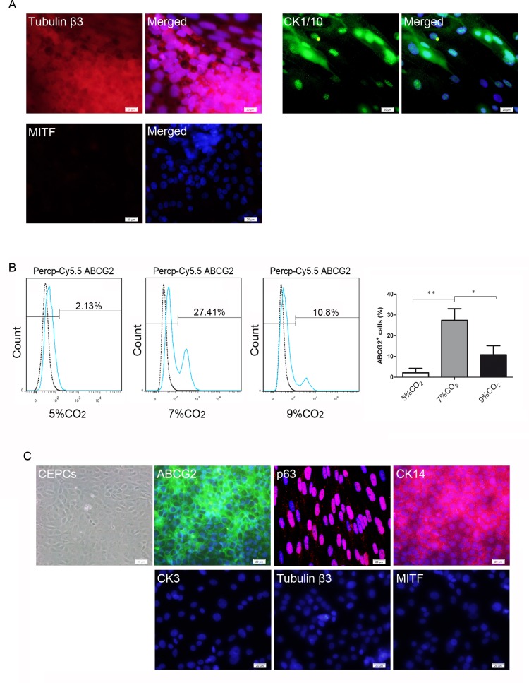
Fig 6. Immunostaining of non-corneal epithelial differentiation markers and isolation of CEPCs derived from hESCs by FACS.
(A) Immunofluorescent staining of tubulin β3, CK1/10, and MITF. (B) FACS analysis of ABCG2-positive cells in different culture conditions at day 6 of differentiation. (C) Immunofluorescent staining of the sorted cells. ABCG2, p63, and CK14 were strongly expressed in the sorted cells. CK3, tubulin β3, and CK1/10 were not detected. Scale bar, 20 μm.

