Figure 6.
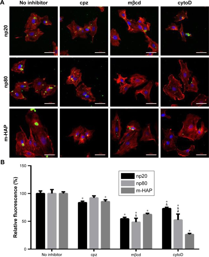
The role of different endocytic pathways in the uptake of HAPs in HUVECs.
Notes: Cells were exposed to HAPs either with or without cpz, mβcd and cytoD. (A) CLSM images of HAP interactions with HUVECs for 2 h. Cells stained for nuclei (blue) and actin (red). HAPs are shown in green. Scale bar: 50 μm. (B) Quantification of the uptake of HAPs after HUVECs were treated with HAPs for 2 h. *P<0.01 versus no inhibitor control, &P<0.01 versus m-HAP group, #P<0.01 versus np20 group.
Abbreviations: CLSM, confocal laser scanning microscopy; cpz, chlorpromazine; cytoD, cytochalasin D; HAP, hydroxyapatite; HUVECs, human umbilical vein endothelial cells; mβcd, methyl-β-cyclodextrin; m-HAP, micro-sized HAP particles.

