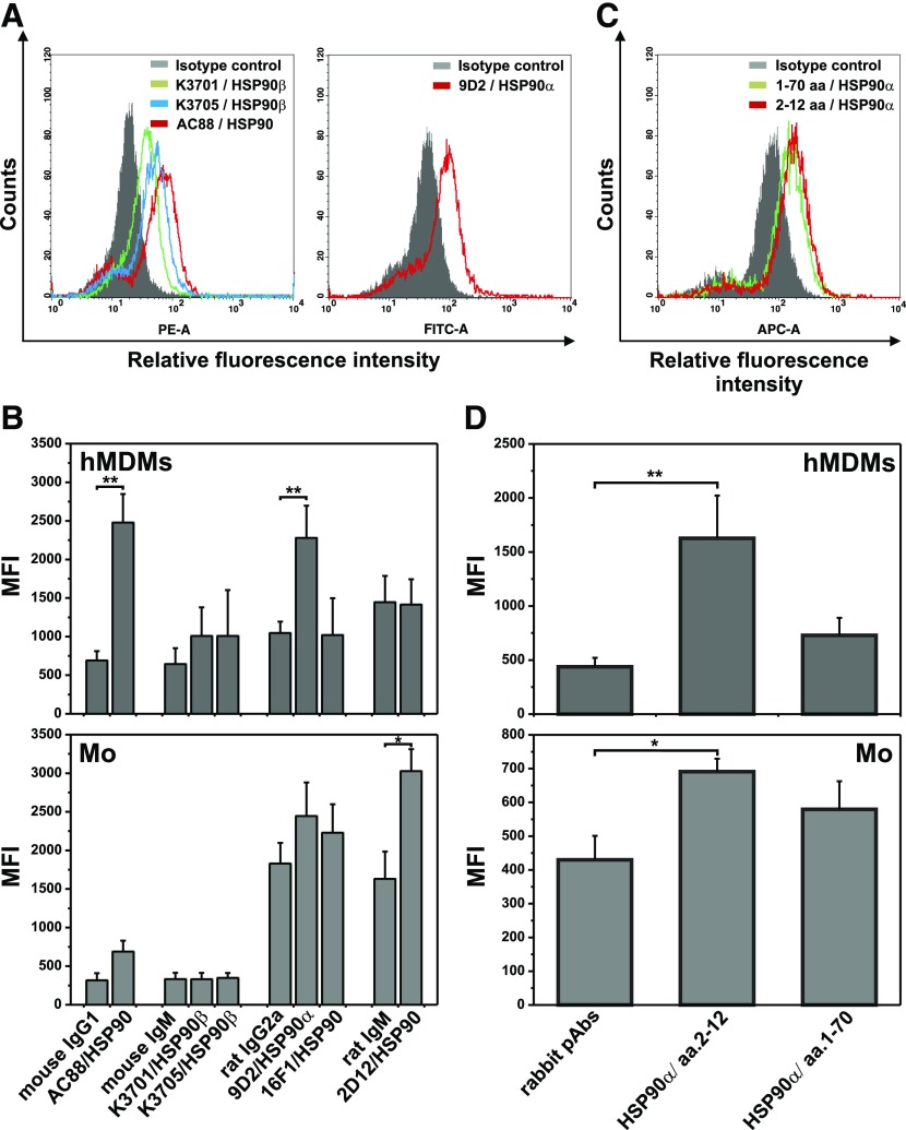
Figure 2. Surface expression of HSP90 on human monocytes and hMDMs.
Monocytes were isolated from PBMC by elutriation. hMDMs were differentiated from adherent monocytes for at least 7 d in medium supplemented with 10% HS and then were nonenzymatically detached. Cells were indirectly labeled with anti-HSP90 mAbs (A and B) (clones denoted in figure captions) or rabbit pAbs (C and D) specific for the N-domain of HSP90 (epitopes denoted in figure captions) and corresponding fluorescent secondary Abs and analyzed by flow cytometry. (A and C) Flow cytometry data from 1 experiment representative of 3 performed showing the expression of HSP90 on hMDMs. The gray-shaded histograms represent background staining obtained with isotype-matched controls. (B and D) HSP90 expression on hMDMs and monocytes, presented as MFI of specific staining in comparison to MFI of corresponding isotype controls (mean ± sem from 3 independent experiments). *P < 0,05; **P < 0.01.

