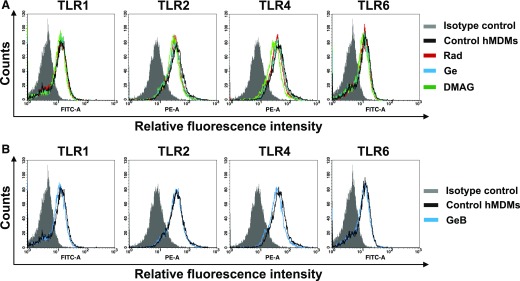
Figure 6. HSP90 inhibitors do not affect surface expression of TLR1, -2, -4, and -6 on hMDMs.
hMDMs left untreated (control) or treated for 4 h with HSP90 inhibitors were nonenzymatically detached, stained with fluorescence-labeled mAbs and analyzed by flow cytometry. (A) Open histograms depict results for control cells or cells treated with Rad or Ge (20 µM) or DMAG (1 µM). The gray-shaded histogram represents background staining obtained with the isotype-matched controls. (B) Open histograms show results for control and cells treated with GeB (20 µM). The gray-shaded histogram represents background staining obtained with isotype-matched controls. Data are from 1 experiment representative of 3 performed.

