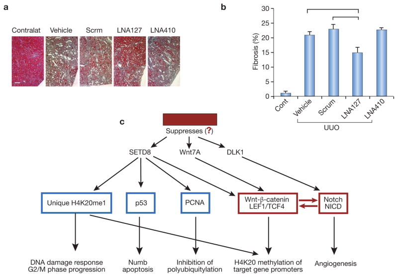
Figure 3. Profibrogenic effect of miR-127-3p.
A and B: Masson’s trichrome staining of UUO kidneys in control mice, mice treated with the negative (scrambled) LNAs, and mice receiving complementary LNAs to miR-127 and miR-410. Ordinate shows the number of pixels corresponding to the detection of fibrotic areas. Horizontal lines above individual bars indicate p<0.05 between the corresponding groups. N=5 per each group of animals. Note that LNA to miR-127 mitigates fibrosis compared to both control groups, whereas LNA to miR-410 (another miR screen finding) is not effective, at least at the dose used.
C: Targets of miR-127-3p. TargetScan identifies these target gene promoters relevant to Wnt/β-catenin and Notch pathways. Note a remarkable overlap with proteomic findings. In red are components of main focus.

