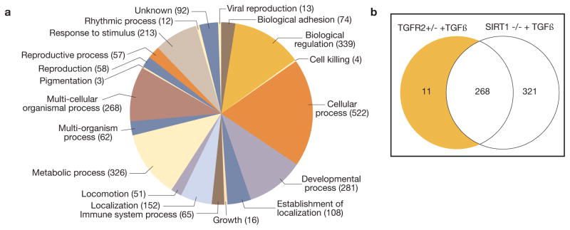
Figure 5.
The secretome of cultured murine renal microvascular endothelial cells. A – categories of proteins detected in the secretome; B – a Venn diagram comparing the protein components of the secretome of endothelial cells isolated from SIRT1−/−, TGFR2+/−, and wild type mice and stimulated with TGF-β1.

