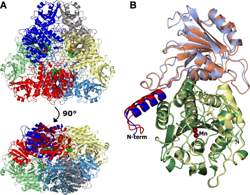
FIG 1 .
Structures of the LAP-As. Protein chains are shown in a ribbon representation. (A) Hexamer of the TbLAP-A--bestatin complex colored by chain. The six bestatin ligands and associated metals are shown as spheres and lie on the inside of the hexamer. (B) Superposition of TbLAP-A and LmLAP-A protomers. The TbLAP-A N-terminal helix (α1) is blue, the N-terminal domain is light blue, and the C-terminal domain is green. For LmLAP-A, α1 is red, the N-terminal domain is coral, and the C-terminal domain is lemon. The Mn2+ ions in the active sites are shown as tan spheres. This and subsequent structure images were made in CCP4mg (63), and superpositions were carried out with SSM (64).

