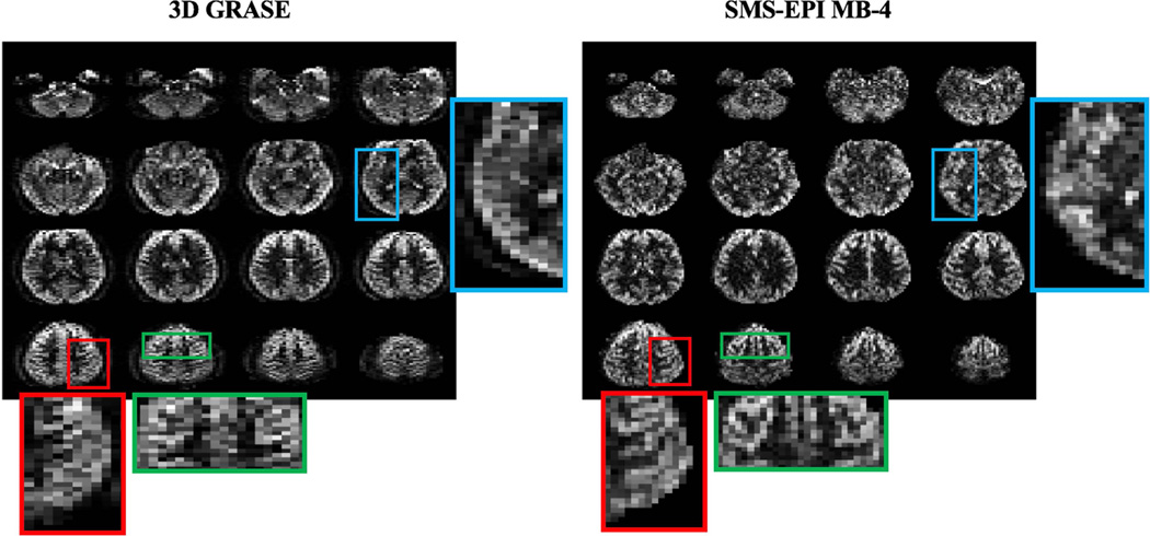
Figure 5.
Perfusion weighted images of central 16 slices from 3D segmented GRASE and SMS-EPI MB-4. For qualitative comparison, even and odd slices in 3D GRASE were added to match the slice position in SMS-EPI scan. Three regions (highlighted by red, green and blue frames) were enlarged for comparison of blurring effects between 3D GRASE and 2D SMS-EPI.

