Figure 4.

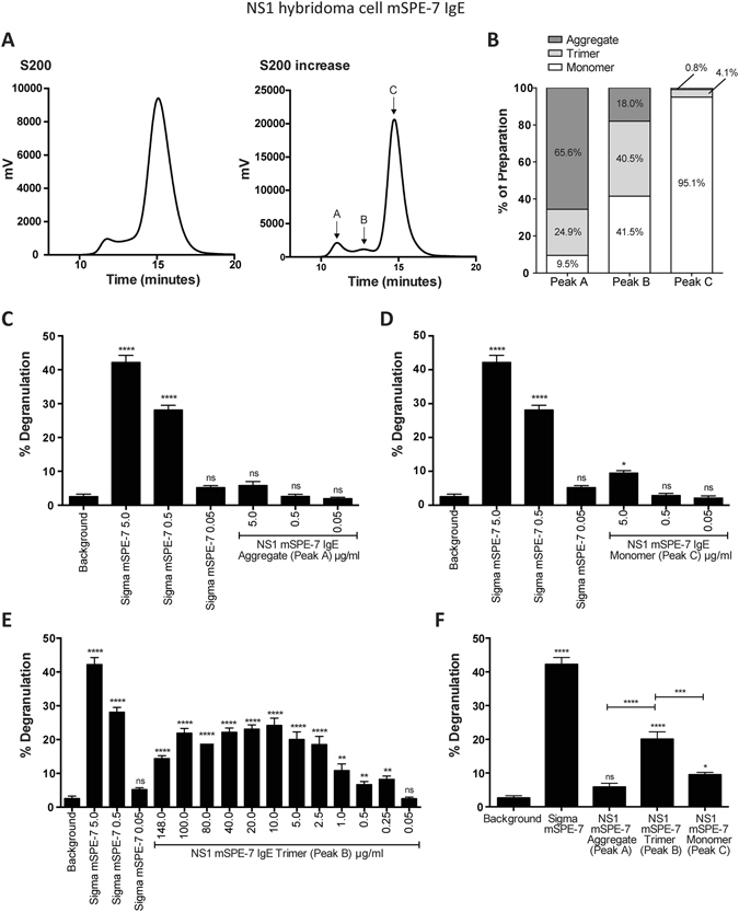
mSPE-7 IgE trimer displays cytokinergic activity. (A) Improved size fractionation of mSPE-7 IgE was achieved with a Superdex 200 Increase HPLC column. (B) SEC-MALLS analysis of peaks A, B and C from the column determined the percentage of high molecular weight aggregates, trimers and monomers in Peaks A, B and C, respectively. The percentages of each in the three fractions are indicated. (C) No RBL-SX38 cell degranulation above buffer background control was induced by incubation with the IgE in peak A preparation (65.6% aggregated IgE), in the absence of antigen. (D) Significant, but low-level degranulation, compared to buffer background control was induced by incubation with 5 μg/ml of the IgE in peak C (95.1% monomeric IgE and 4.1% trimeric IgE) in the absence of antigen. Incubation with unpurified Sigma mSPE-7 IgE, in absence of antigen, resulted in significant RBL-SX38 degranulation compared to buffer background control in both experiments. (E) Significant RBL-SX38 cell degranulation was induced by incubation with NS1 mSPE-7 IgE trimer in the absence of antigen (peak B in purification profile). (F) Degranulation induced by peaks A, B and C at 5 μg/ml is compared. Means of 3 independent experiments ± SEM are shown. Statistically significant difference to background control (unless otherwise indicated) was determined by one-way ANOVA with Dunnett’s post-test; ****P < 0.0001, **P = 0.001 to 0.01, ns P > 0.05.
