
Figure 3.
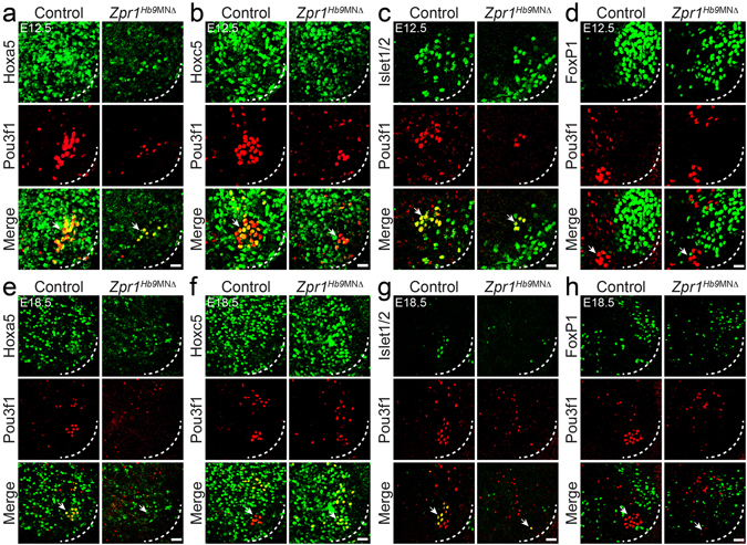
ZPR1 deficiency causes progressive loss of PMC neurons in Zpr1 Hb9MNΔ mice. Spinal cord sections of the cervical region (C3-C5) from (a–d) E12.5 and (e–h) E18.5 control and Zpr1 Hb9MNΔ embryos were stained with antibodies against Pou3f1, Hoxa5, Hoxc5, Islet1/2 and FoxP1 proteins. Immunofluorescence images reveal the effect of Zpr1 mutation on the loss and disorganization of different populations of neurons in the cervical region, including PMC and LMC groups of motor neurons. Quantification of Pou3f1+, Hoxa5+, Hoxc5+, and FoxP1+ neurons at E12.5 and E18.5 stages is presented in Supplementary Figure S3. Neurons were counted in serial sections of C3-C5 region. Neuron losses are presented as (mean ± s.e.m.; n = 3 mice/group). At E12.5 stage, images (a–d) and quantification shows loss (64.29 ± 4.78%, p < 0.0001) of Pou3f1+ motor neurons. By E18.5 stage (e–h), Pou3f1+ neurons were barely detectable and quantification shows greater loss (90.00 ± 15.81%, p = 0.0013) of neurons in Zpr1 Hb9MNΔ embryos. Image analysis and quantification of Hoxa5+ neurons shows non-progressive loss of neurons at E12.5 (64.64 ± 15.54%, p = 0.0032) and at E18.5 (59.35 ± 11.60%, p = 0.0022) stages in Zpr1 Hb9MNΔ embryos. A smaller loss of Hoxc5+ neurons at E12.5 (31.93 ± 4.80%, p = 0.0002) and E18.5 (24.51 ± 9.81%, p = 0.0369) stages was noted in Zpr1 Hb9MNΔ mice. A relatively smallest decrease in FoxP1+ LMC neurons at E12.5 (13.59 ± 10.94%, p = 0.2605) that increases to a statistically significant loss at E18.5 (28.17 ± 2.42%, p = 0.0001) was found in Zpr1 Hb9MNΔ mice. Images (d,h) show disorganization and the loss of compactness of FoxP1+ LMC neurons. Dotted arcs represent the orientation of view from the anterior horn side of the spinal cord. Arrows indicate phrenic nerve motor neurons. Scale bars are 25 μm (a–d) and 50 μm (e–h).