Figure 4.

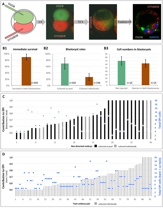
Tracing of the contribution of sister two-cell stage blastomeres to epiblast. Each blastomere in two-cell embryos were microinjected with fluorescent dextran beads of different colors (OGDB, OY568DB), followed by in vitro culture to blastocyst and NANOG immunofluorescence analysis (A). The quality of the microinjections was validated by survival rates of the blastomeres, blastocyst rates, and total cell numbers of blastocysts (B). The relative contribution of each two-cell blastomere to EPI (ratio from 0.5 to 1) and the absolute total number of EPI cells (•) are shown for non-bisected (C) and twin (D) embryos. Error bars in (B) are standard deviations. Black and gray bars in (C) and (D) indicate embryos cultured as pools and embryos cultured individually, respectively.
