
Figure 1.
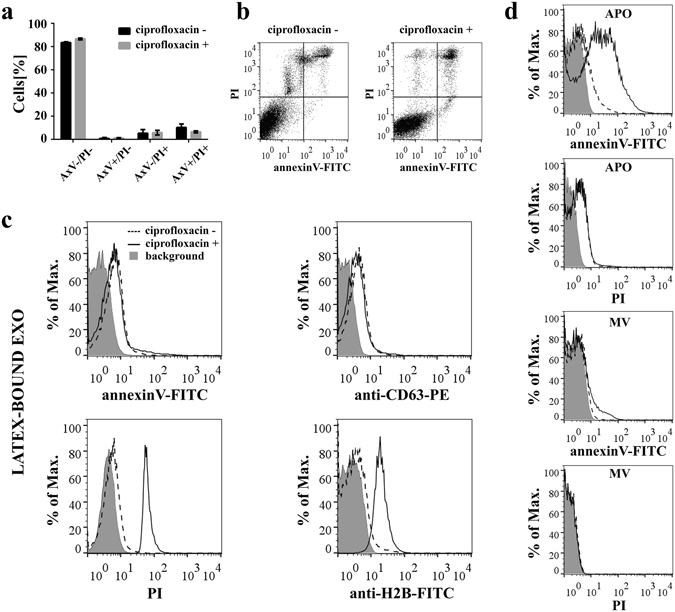
Effects of sustained ciprofloxacin exposure on Jurkat cells. (a,b) Viability of Jurkat cells with/without exposure to ciprofloxacin (10 µg/mL for >14 days) was analyzed by flow cytometry after staining with annexinV-FITC and propidium iodide (PI). (a) Mean values+/− S.D. (error bars) of two independent experiments are shown in the histogram plot. AxV: annexinV. (b) Representative dot plots showing the four quadrants of annexinV-FITC and PI stained Jurkat cells. (c) Exosomes (EXOs) derived from ciprofloxacin-exposed/unexposed Jurkat cells were conjugated onto latex beads and characterized by flow cytometry after staining with annexinV-FITC, anti-CD63-PE, PI or anti-histone H2B-FITC. The background fluorescence of stained latex beads is indicated by grey histograms. (d) Microvesicles (MVs) and apoptotic bodies (APOs) were labeled directly with annexinV-FITC and PI. The background fluorescence of EVs is indicated by grey histograms.