
Figure 5.
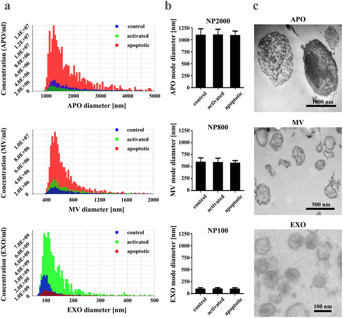
Characterization of extracellular vesicles (EVs) released by ciprofloxacin-exposed control, activated or apoptotic Jurkat cells. (a) Concentration values of EVs in 100 µL (isolated from the supernatant of 2.5 × 10⁷ cells) were determined by tunable resistive pulse sensing (TRPS), and are plotted as a function of their diameter for control, activated or apoptotic cells (blue, green or red colors, respectively). Histogram plots of EVs are representative of three independent experiments. (b) Mode diameters of EV subpopulations were determined by TRPS, and are presented+/− S.D. (error bars) of three independent experiments, for control, activated and apoptotic cells. NP2000, NP800 and NP100 IZON nanopore membranes were used. (c) Transmission electron microscopy images of apoptotic bodies (APOs), microvesicles (MVs) and exosomes (EXOs) samples.