
Figure 6.
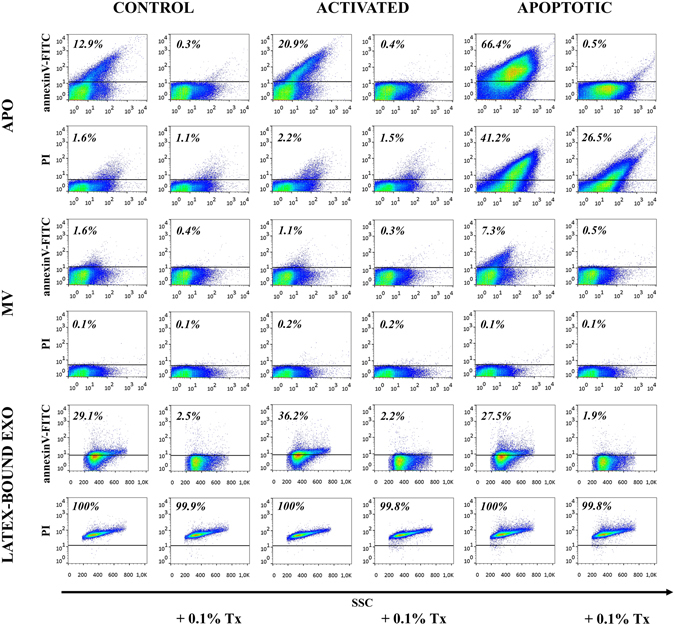
Flow cytometry analysis of extracellular vesicles (EVs) derived from ciprofloxacin-exposed control, activated or apoptotic Jurkat cells. EVs were stained by annexinV-FITC and propidium iodide (PI) for flow cytometry and analyzed before and after detergent lysis with 0.1% Triton X-100. Apoptotic bodies (APOs) and microvesicles (MVs) were labeled and measured directly, whereas exosomes (EXOs) were conjugated onto latex beads before staining. Density plots show annexinV-FITC and PI positivity of EVs derived from ciprofloxacin-exposed control, activated and apoptotic Jurkat cells. The percentages of positive events (above the threshold represented by a black line) are shown in the plots.