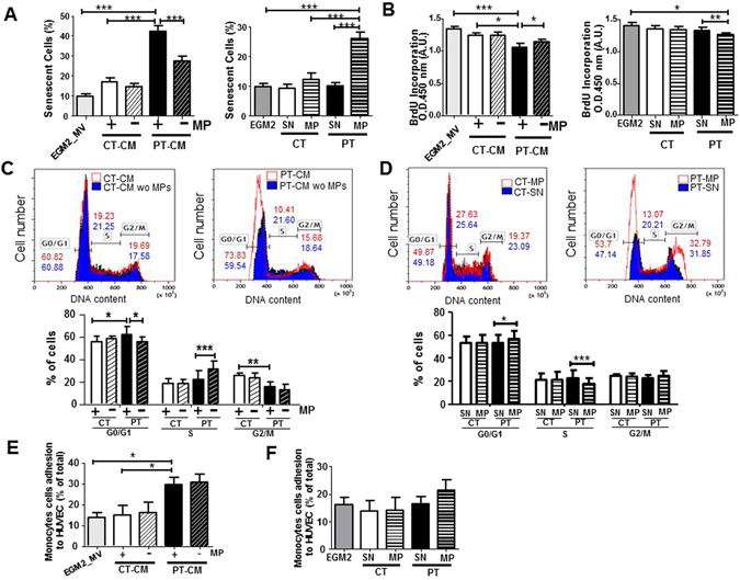
Figure 2.

EMP components of the PT-SASP are key mediators of paracrine senescence. (A) Quantification of positive senescence-associated (SA) β-galactosidase staining in HUVEC exposed for 48 h to conditioned media (CM) ± depleted in EMP (left) or vehicle (SN), CT-EMP or PT-EMP (right). (HUVEC, N = 4; N = 6 CT vs. 7 PT). (B) Proliferation was assessed in a BrdU incorporation assay. The impact of CM ± depleted in EMP (left). Impact of purified EMP (right). (HUVEC, N = 4; N = 6 CT vs. 7–9 PT). (C,D) Cell cycle distribution by flow cytometry. DNA content in each cell cycle phase in HUVEC was analyzed by flow cytometry after propidium iodide staining. Representative histograms (upper panel) are from HUVEC treated with CM ± depleted in EMP (C) or purified EMP from CT- and PT-ECFC (D). Graph (lower panel) represents a mean percentage of cells at different phases of the cell cycle determined by the DNA content ± SD for 4 independent HUVEC treated with 6 CT vs 7–9 PT samples. *p < 0.05; **p < 0.01; ***p < 0.001 (E,F) THP1 monocytic cell line adhesion assay on HUVEC exposed overnight to (E) CM ± depleted in EMP or (F) vehicle (SN), CT-EMP or PT-EMP. Data are represented as means ± SEM for 4 independent HUVEC treated with 4 CT vs 7 PT independent samples. Each experiment was carried out in triplicate. *p < 0.05.
